Surgical and endodontic management of autotransplantation of third molars in adults
Introduction
The aim of our research was to observe the formation periodontal ligament after autotransplantation of third molars with closed tip in adults. The quality of reattachment depends on many factors such as the amount of saved fibers on roots and bone, or the width of the alveolar defect. In this study, we want to determine how the ligament is formed depending on the state of the recipient zone, where the periodontal ligament of bone was strongly reduced, the ligament stayed untouched, and where there was no ligament at all.
Materials and methods
In the study we observe 50 patients who did undergo autotransplantation of third molars with a closed root tip. All clinical cases are divided into three groups. The 1st group is presented by autotransplantation in a fresh socket without signs of inflammation. The 2nd group - in a fresh socket, with the presence of a chronic inflammatory process and in the 3rd group the socket was prepared in native bone. The transplanted teeth were endodontically treated before or in 14 days after the operation. After removing a splint (the average time of splinting is 30 days) the Periotest measurements were done. All autotransplanted teeth were put in the occlusion.
Results
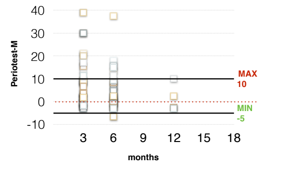
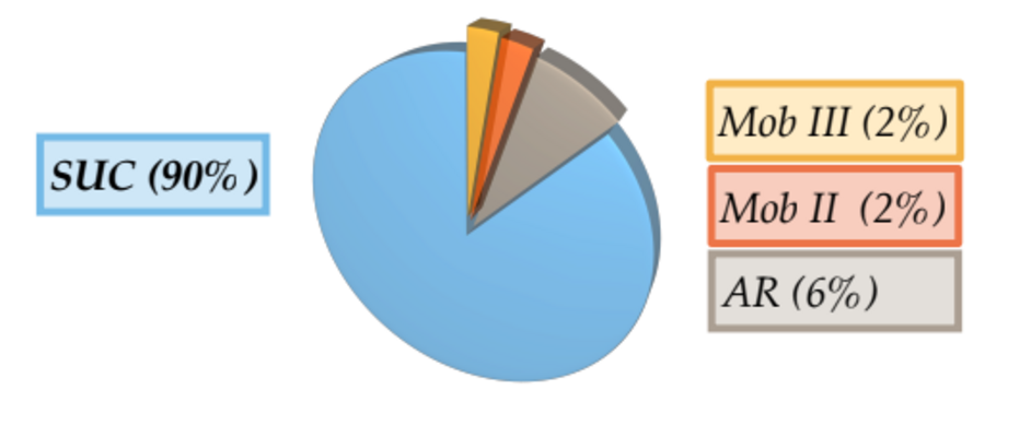
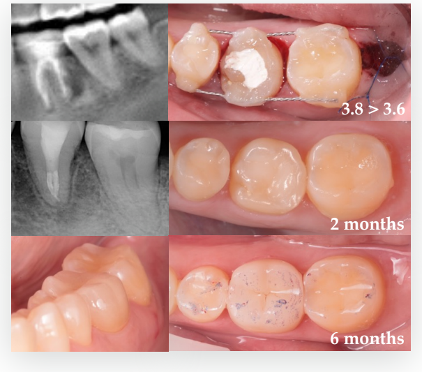
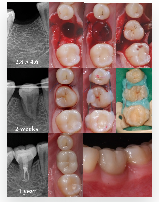
The first results were evaluated after 3 and 6 months. mobility was 36 (72%), I – 9 (18%), II – 4 (8%) and III – 1 (2%), AR – 0. After 6 months, 41 teeth had normal mobility (82%), I – 4 (8%), II – 1 (2%), III - 1 (2%), AR – 3 (6%). The depth of periodontal attachment did not exceed 3 mm, but in 8 cases (16%) there were recessions according to I Miller class. These parameters were stable from 3 till 6 months. During the X-ray examination, it was found after 3 months that in 26 (52%) cases, PDL did not differ significantly from the PDL of adjacent teeth. In 15 (30%) cases PDL had an uneven narrow structure, less than 1 mm thick. In 3 cases (6%), signs of replacement resorption were found and in 6 (12%) PDL expansion was observed. After 6 months in 41 (82%) cases there were no signs of PDL abnormality, excepting the thinness.
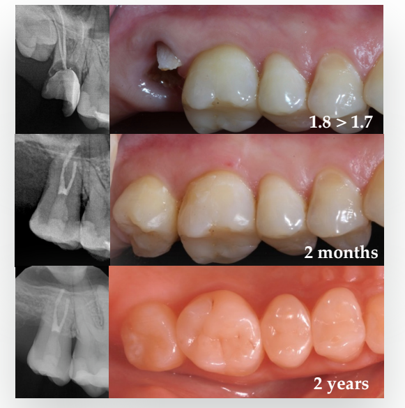
Case #1 (1st group)
Case #2 (2nd group)
Case #3 (3rd group)
Conclusions
Since the study is still ongoing, we can evaluate the first results of autotransplantation after 3 and 6 months and draw preliminary conclusions. Using Periotest control, periodontal probing, and regular x-ray examination, we are able estimate the PDL state sufficiently. It was found that the phenomenon of replacement resorption can not be evaluate earlier than 6 months after the transplantation. We also determined that the gingival attachment that had firstly formed does not tend to change. It has also been shown that despite the thinness of PDL on the radiograph, it does not differ functionally from other teeth.
Authors: Badalyan Kristina, Zedgenidze Alena
Literature cited
- Schwartz O1, Andreasen JO. J Oral Maxillofac Surg. 1988 Aug;46(8):672-81. Allotransplantation and autotransplantation of mature teeth in monkeys: the influence of endodontic treatment.
- Andreasen JO1, Paulsen HU, Yu Z, Schwartz O. Eur J Orthod. 1990 Feb; 12(1):14-24. A long-term study of 370 autotransplanted premolars. Part III. Periodontal healing subsequent to transplantation.
- Yu HJ1, Jia P1, Lv Z1, Qiu L. Int J Oral Maxillofac Surg. 2017 Apr;46(4): 531-538. Epub 2017 Jan 3. Autotransplantation of third molars with completely formed roots into surgically created sockets and fresh extraction sockets: a 10-year comparative study.