Clinical experience of using an individual piezosurgical tip for tooth autotransplantation into native bone.
Abstract
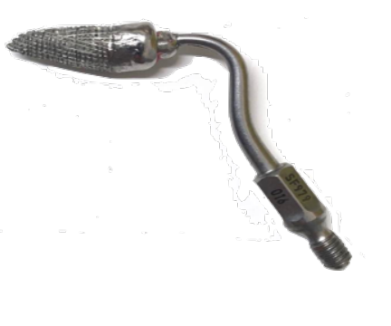
In this study, a wisdom tooth was transplanted into the native bone. This type of operation is usually complicated by the creation of a socket of a suitable size. We propose a method to create a socket the same form as a donor tooth. To create a congruent socket we use a custom-made piezosurgical tip made of a medical cobalt-chromium alloy which was done with 3D model. The tip was sharpen and glued to a standard cap of a piezosurgical hand-piece. After creating a socket, the tooth was autotransplanted. The endodontic treatment was initiated in two weeks and the crown restoration was made in six months after the surgery.
Clinical case
A 25-years-old patient, with no medical history of interest attended to install an implant to the missing 16. During the CBCT-scan analysis we determined the most optimal position of the transplanted tooth in recipient region so we glued the piezosurgical tip in a way according to these measurements.
Operation process
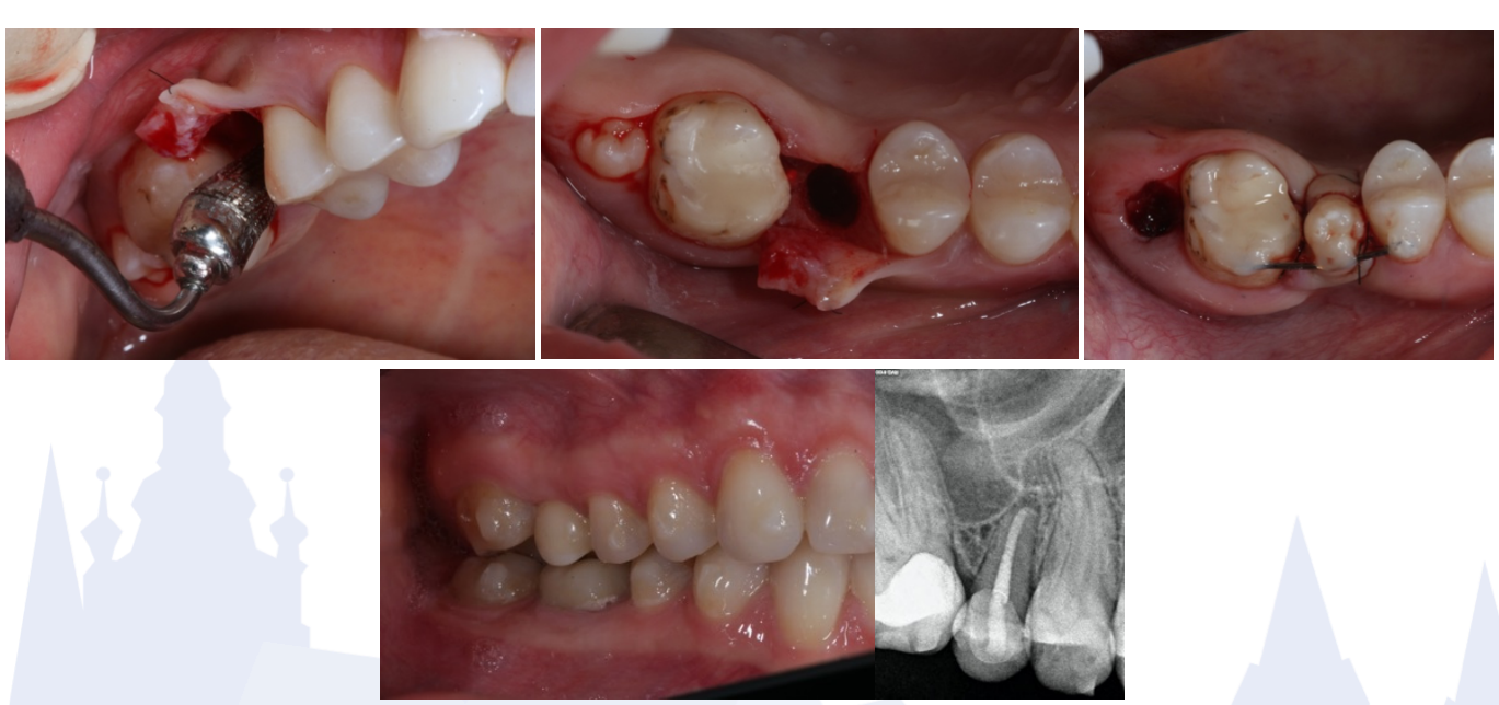
The cortical bone was drilled with a ball-shaped surgical bur, then the individual piezosurgery tip was used. A congruent socket was formed and tooth 18 was carefully removed and moved into the prepared socket. The extra-alveolar time was less than 30 seconds. The tooth was fixed with a ligature. The soft tissues are sutured.
Postoperative period
The sutures have been removed after seven days. Endodontic treatment of the transplanted tooth was performed after two weeks. The ligature was removed tree weeks after the operation. Six months after the operation, periotestometry data was +16 (1.6) and +1.2 (2.6), respectively. Then the tooth was covered with ceramic crown. After 2 years the tooth had no signs of resorption and periapical inflammation. Periotestometry data was +9.9, which corresponds to normal mobility.
Discussion
The use of an individual piezosurgical tip allows to form a recipient hole corresponding with shape and size to the transplanted tooth. This method avoids bone pressure on the root and at the same time the distance between these surfaces is minimal. This approach reduces the risk of resorption, reduces the healing time. It also greatly facilitates the work of the surgeon and reduces the risk of postoperative complications.